
A pulmonary embolism (PE) — commonly called a lung embolism — occurs when a blood clot (usually from the deep veins of the legs) breaks free, travels through the bloodstream, and lodges in the arteries supplying the lungs. It blocks blood flow, strains the heart, and can drop oxygen levels rapidly.
It is not rare. Pulmonary embolism is the third most common cause of cardiovascular death worldwide, after heart attack and stroke. In the United States alone, an estimated 300,000–600,000 people are affected each year. What makes it particularly dangerous is that its symptoms overlap with dozens of less serious conditions — and even experienced clinicians sometimes miss it on first presentation.
This guide walks you through every key warning sign, what causes lung embolism, who is most at risk, how it’s diagnosed, and what the treatment involves. Read it, share it, and know it before you need it.
What Is a Pulmonary Embolism (Lung Embolism)?
The lungs have a dense network of blood vessels. Oxygenation depends on constant, unobstructed blood flow through these vessels. When a clot — known medically as a thrombus — breaks away from its original location (most commonly the deep veins of the calf, thigh, or pelvis) and travels to the lung, it blocks one or more of these vessels.
This blockage does two dangerous things simultaneously: it prevents part of the lung from exchanging oxygen and carbon dioxide, and it forces the right side of the heart to work against dangerously elevated resistance. If the clot is large enough, the right ventricle can fail within minutes.
A pulmonary embolism is a blockage in the lung’s arteries caused by a blood clot that travels from elsewhere in the body — usually the deep veins of the legs. It reduces oxygen levels, strains the heart, and is life-threatening if not treated rapidly. It is a medical emergency requiring immediate hospital care.
The Clot Journey: From Leg Vein to Lung Artery
Most pulmonary emboli originate as deep vein thrombosis (DVT) — a clot in the deep veins of the lower leg or thigh. These clots form when blood pools or moves too slowly, often due to prolonged immobility, vein injury, or a clotting disorder.
Once a DVT clot dislodges, it travels up through the inferior vena cava, enters the right side of the heart, and gets pumped directly into the pulmonary arteries. The narrowing arteries trap the clot, and the blockage begins.
Signs of Lung Embolism: The Full Symptom Breakdown
The signs of lung embolism exist on a spectrum. They depend on the size of the clot, how many vessels are blocked, and the person’s baseline cardiovascular health. A small clot in a healthy person may cause mild breathlessness. A massive embolism can cause sudden cardiac arrest with no warning at all.
Understanding the range of symptoms — from subtle to critical — is what separates early treatment from a missed diagnosis.
The Classic Triad (Textbook Presentation)
Medical education has long taught the classic triad of pulmonary embolism symptoms. It is worth knowing — though it’s important to understand that fewer than 30% of patients present with all three together:
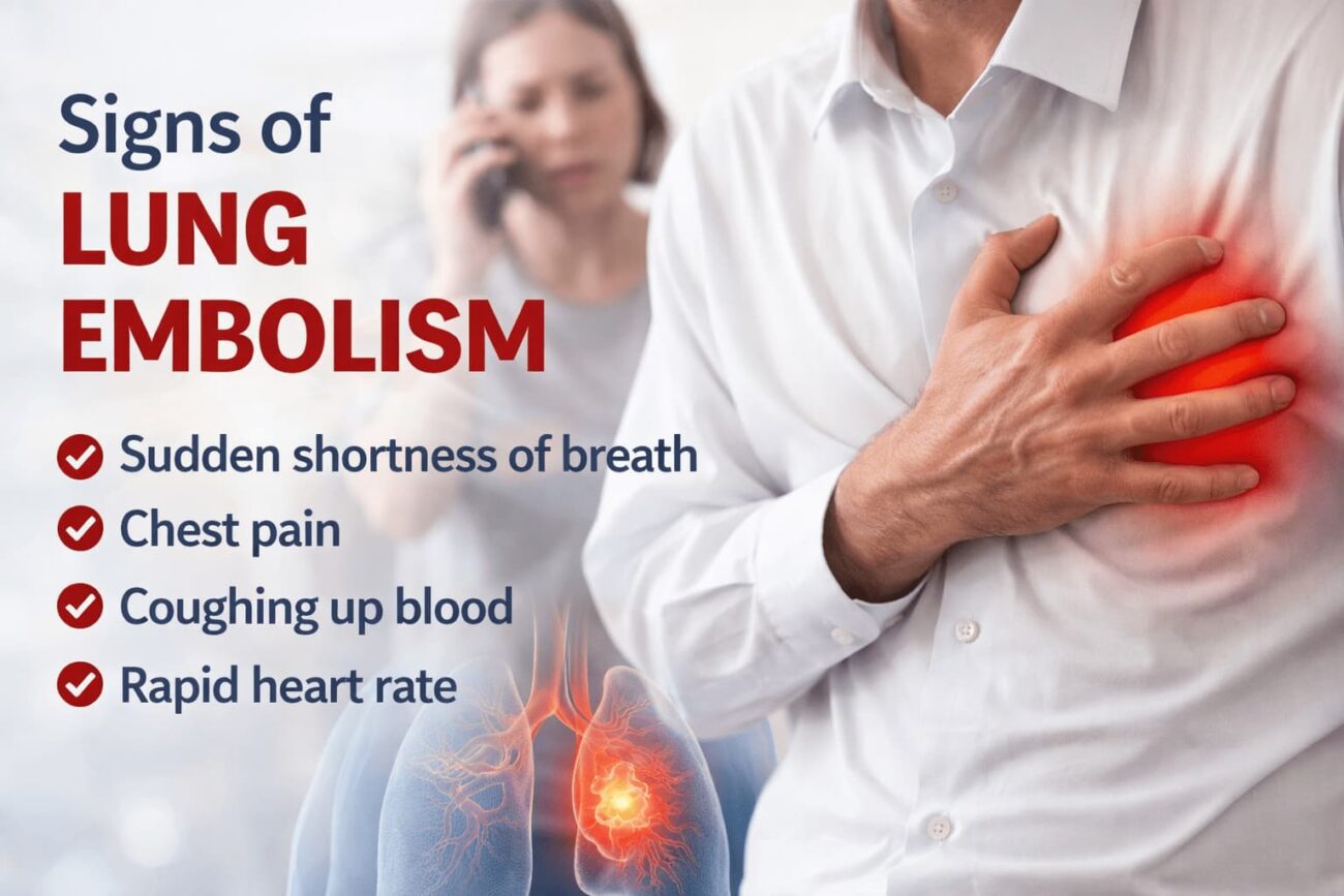
- Sudden shortness of breath — especially at rest or with minimal exertion
- Pleuritic chest pain — sharp pain that worsens when you inhale deeply
- Hemoptysis — coughing up blood or blood-streaked mucus
Full Range of Pulmonary Embolism Symptoms
- Sudden, unexplained shortness of breath — the most common symptom, present in ~80% of cases
- Sharp chest pain — often on one side, worsens with deep breathing or coughing
- Rapid heart rate (tachycardia) — the heart accelerating to compensate for low oxygen
- Coughing, sometimes with blood — especially if a small lung infarction (tissue death) has occurred
- Dizziness or lightheadedness — from reduced oxygen delivery to the brain
- Excessive sweating — cold, clammy skin is a stress response
- Anxiety or sense of impending doom — a frequently dismissed symptom that clinicians now take seriously
- Low-grade fever — from the inflammatory response to clotted tissue
- Swollen, painful leg — a sign of the underlying DVT that caused the embolism
- Bluish discoloration of the lips or fingertips (cyanosis) — indicates severe oxygen deprivation
- Fainting or collapse — a sign of massive PE and hemodynamic instability
When Symptoms Are Subtle or Atypical
Not every pulmonary embolism announces itself dramatically. Small or subsegmental PEs can cause only mild, persistent breathlessness — the kind that patients attribute to being unfit, anxious, or fighting a chest infection. Some people notice they’re simply unable to take a full, satisfying breath.
Older adults and people with pre-existing lung or heart conditions may have atypical presentations. Their baseline symptoms can mask what a PE adds on top. In these cases, a clinical suspicion score — combined with a D-dimer blood test and imaging — is essential.
Risk Factors: Who Is Most Likely to Get a Lung Embolism?
Understanding your personal risk is not just an academic exercise — it changes how urgently you should seek evaluation when borderline symptoms appear. The more risk factors you carry, the lower your threshold should be for calling a doctor or going to the emergency room.
Virchow’s Triad: The Root Cause Framework
Medical science has understood since the 19th century that blood clots form when three things converge — a framework called Virchow’s Triad:
- Slowed blood flow — from immobility, obesity, heart failure
- Damage to blood vessel walls — from surgery, trauma, or IV catheters
- Increased clotting tendency (hypercoagulability) — from genetic disorders, pregnancy, oral contraceptives, or cancer
Specific Risk Factors for Pulmonary Embolism
| Risk Category | Examples | Relative Risk Level |
|---|---|---|
| Recent surgery | Hip/knee replacement, major abdominal surgery | 🔴 Very High |
| Prolonged immobility | Long-haul flights, bed rest, hospitalization | 🔴 High |
| Active cancer | Especially pancreatic, lung, and brain cancers | 🔴 Very High |
| Previous VTE | Prior DVT or PE in personal history | 🔴 Very High |
| Inherited clotting disorders | Factor V Leiden, Protein C/S deficiency, antiphospholipid syndrome | 🔴 High |
| Hormonal therapy | Combined oral contraceptive pill, HRT | 🟠 Moderate |
| Pregnancy and postpartum | Risk is highest in the 6 weeks after delivery | 🔴 High |
| Obesity | BMI over 30 significantly raises DVT risk | 🟠 Moderate |
| Heart failure or stroke | Reduced cardiac output slows venous return | 🟠 Moderate |
| Smoking | Damages vessel walls, promotes clotting | 🟡 Low–Moderate |
| Dehydration | Thickens blood and slows flow | 🟡 Low–Moderate |
How Pulmonary Embolism Is Classified by Severity
Not all pulmonary emboli are equal. Clinicians classify them by hemodynamic impact — how significantly they compromise the heart and blood pressure — because this determines treatment urgency and approach.
Low-Risk (Subsegmental) PE
Small clots in peripheral lung vessels. Often found incidentally on CT scans. Patients are hemodynamically stable with minimal symptoms.
Submassive (Intermediate) PE
Clot is larger and shows early signs of right heart strain on imaging or troponin tests. Blood pressure remains stable but the situation can deteriorate.
Massive PE
Severe right ventricular failure. Systolic blood pressure drops below 90 mmHg. High immediate mortality risk. Requires aggressive treatment — thrombolytics or surgery.
How Lung Embolism Is Diagnosed
Diagnosing a pulmonary embolism requires a structured clinical approach. No single test confirms it — instead, clinicians use a combination of probability assessment, blood markers, and imaging.
Step 1: Clinical Probability Scoring
The Wells PE Score is the most widely used pre-test probability tool. It scores points for factors like heart rate above 100, signs of DVT, no alternative diagnosis being more likely, and prior PE or DVT history. The total score guides how aggressively to investigate.
Step 2: D-Dimer Blood Test
D-dimer is a protein fragment released when a blood clot dissolves. A negative D-dimer in a low-probability patient effectively rules out PE. However, D-dimer is non-specific — it rises in infection, pregnancy, surgery, and even stress — so a positive result alone does not confirm PE.
Step 3: CT Pulmonary Angiography (CTPA)
CTPA is the gold standard imaging test for pulmonary embolism. A contrast dye is injected into a vein, and CT imaging maps the pulmonary arteries in detail. Clots appear as filling defects. CTPA can detect clots down to subsegmental level and takes only minutes to perform in most hospitals.
Additional Diagnostic Tools
- Chest X-ray: Usually normal in PE, but rules out other causes of chest pain and breathlessness (e.g., pneumonia, pneumothorax)
- ECG (electrocardiogram): May show right heart strain patterns — the classic “S1Q3T3” pattern or sinus tachycardia
- Echocardiogram: Assesses right ventricular function in suspected massive PE
- V/Q scan (ventilation-perfusion scan): Used when CTPA is contraindicated (e.g., severe kidney disease or contrast allergy)
- Troponin and BNP: Elevated levels indicate right heart strain and predict higher risk of deterioration
- Compression ultrasound of legs: Detects DVT that may have seeded the PE
Treatment Options for Pulmonary Embolism
Treatment depends on severity. The two primary goals are: preventing the existing clot from growing, and stopping new clots from forming. In life-threatening cases, rapid clot dissolution becomes the priority.
Anticoagulation Therapy (The Cornerstone)
For most patients with confirmed PE, anticoagulant therapy is the first and primary treatment. It does not dissolve the clot directly — the body’s own fibrinolytic system does that over weeks — but it prevents the clot from extending and blocks new ones from forming.
- DOACs (Direct Oral Anticoagulants) — rivaroxaban, apixaban, edoxaban — are now first-line therapy for most patients. They are taken by mouth, require no routine blood monitoring, and have a predictable effect.
- Low Molecular Weight Heparin (LMWH) — injected subcutaneously; preferred in pregnancy and cancer-related VTE.
- Unfractionated Heparin (IV) — used in massive PE and when rapid reversal may be needed (e.g., pre-surgery).
- Warfarin — older oral anticoagulant; still used when DOACs are contraindicated or cost-prohibitive. Requires regular INR monitoring.
Thrombolysis (Clot-Busting Therapy)
In massive PE with cardiovascular collapse, systemic thrombolysis using drugs like alteplase (tPA) can rapidly dissolve the clot and restore pulmonary blood flow. The benefit is fast — but the risk of major bleeding, including brain hemorrhage, is significant. It is reserved for patients in imminent risk of death.
Catheter-directed thrombolysis — delivering lower doses of thrombolytics directly to the clot via a catheter — is an emerging option that reduces systemic bleeding risk.
Surgical Embolectomy
In rare cases where thrombolysis has failed or is absolutely contraindicated, open-chest surgical removal of the clot can be life-saving. It is a last-resort procedure with significant operative risk.
IVC Filter
An inferior vena cava (IVC) filter is a small, umbrella-like device placed in the main vein returning blood to the heart. It physically traps clots traveling from the legs before they reach the lungs. IVC filters are used when anticoagulation is contraindicated — for example, in patients at very high bleeding risk.
PE vs. Panic Attack vs. Heart Attack: How to Tell the Difference
Because PE symptoms overlap significantly with panic attacks, anxiety disorders, and acute coronary syndrome (heart attack), misattribution is common — and dangerous. Here’s how they typically differ:
| Feature | Pulmonary Embolism | Panic Attack | Heart Attack |
|---|---|---|---|
| Onset | Sudden, often at rest | Sudden, often triggered by stress | Sudden or gradual onset |
| Chest pain type | Sharp, worsens with breathing | Tight, pressured, often central | Crushing, squeezing, may radiate to arm or jaw |
| Oxygen saturation | Often low (SpO₂ <95%) | Normal | May drop in severe cases |
| Coughing blood | Possible | No | No |
| Leg swelling or pain | Common (from underlying DVT) | No | No |
| ECG changes | Right heart strain pattern | Normal or mild sinus tachycardia | ST elevation or depression |
| Resolves with breathing exercises | No | Often yes | No |
When in doubt, go to the emergency room. A false alarm costs you a few hours and some discomfort. Ignoring a real PE can cost you your life.
Preventing Lung Embolism: Evidence-Based Strategies
Prevention is most relevant for people with known risk factors — especially before and after surgery, during long travel, and in medical illness requiring bed rest.
During Hospitalization or Post-Surgery
- Pharmacological prophylaxis: Low-dose LMWH or aspirin prescribed by your surgical or medical team reduces DVT risk significantly.
- Compression stockings: Graduated compression stockings maintain venous flow in immobile patients.
- Intermittent pneumatic compression (IPC) devices: These inflatable leg sleeves are standard in post-operative patients.
- Early mobilization: Getting patients up and walking as soon as medically possible is one of the most effective PE prevention strategies known.
During Long-Distance Travel
- Stand and walk every 1–2 hours on long flights or road trips
- Do in-seat calf raises and ankle circles regularly
- Stay well hydrated — dehydration thickens blood
- Consider compression travel socks if you have risk factors
- Discuss aspirin or LMWH prophylaxis with your doctor if you have prior VTE history
Long-Term Lifestyle Factors
Maintaining a healthy weight, staying physically active, and avoiding prolonged sitting significantly reduce baseline clot risk. For evidence-based guidance on managing weight-related metabolic risks, see our guide on evidence-based weight management strategies Internal.
It is also worth noting that micronutrient deficiencies can influence cardiovascular health and inflammatory pathways that contribute to clotting risk. For a broader picture of how nutrient status affects your circulatory system, read about the warning signs of magnesium deficiency and cardiovascular health Internal.
Signs of Lung Embolism in Special Populations
Pregnancy and Postpartum
Pregnancy creates a state of natural hypercoagulability — necessary to prevent excessive bleeding during childbirth. This same mechanism, however, raises the risk of DVT and PE throughout pregnancy and for up to 12 weeks postpartum. PE is one of the leading causes of maternal death in high-income countries.
Symptoms of PE in pregnancy — breathlessness and rapid heart rate — are common in normal pregnancy, making diagnosis especially challenging. Any pregnant woman with sudden or worsening unexplained breathlessness, chest pain, or unexplained tachycardia should be evaluated urgently.
Cancer Patients
Cancer is one of the strongest independent risk factors for VTE. Certain cancer types (pancreatic, brain, gastric, lung) are particularly thrombogenic. Chemotherapy, central venous catheters, and immobility further multiply risk. Many oncology centers now provide routine VTE prophylaxis protocols for high-risk patients.
Elderly Patients
Older adults often present with atypical or minimal symptoms. Confusion, unexplained deterioration in mobility, or sudden functional decline may be the only clue in elderly patients. Comorbidities including heart failure and COPD complicate interpretation of breathlessness.
What Major Medical Guidelines Recommend
Pulmonary embolism management is governed by well-established international guidelines. The European Society of Cardiology (ESC) and the American Heart Association (AHA) both publish regularly updated clinical practice guidelines on VTE diagnosis and management.
The ESC’s guidelines emphasize a risk-stratification-based approach — treatment intensity should match clinical severity. The full guidance is available at the ESC Clinical Practice Guidelines on Acute Pulmonary Embolism.
The Centers for Disease Control and Prevention (CDC) also maintains a publicly accessible resource on blood clot awareness at CDC.gov’s DVT and PE information page, which is an important educational reference for patients and caregivers.
For individuals who want to monitor overall vascular and metabolic health — including reading related guides on inflammation, gut health, and supplements that affect cardiovascular risk — explore the Symptoms & Health Conditions section at HealthMetricLab Internal.
And if you’ve been prescribed anticoagulants long-term and are also managing related health conditions involving liver function or antiviral therapy, our detailed resource on antiviral medication interactions and liver health Internal may be relevant to your overall care picture.
Frequently Asked Questions About Signs of Lung Embolism
Yes — and this is why PE is so commonly missed. Shortness of breath alone, unexplained rapid heart rate, or sudden fatigue can each be the presenting sign of a PE without any chest pain whatsoever. Some patients, particularly the elderly or those with large reserves, may only notice they “can’t get a full breath” for a few days before seeking help.
Small, peripheral PEs can go undetected for days or even weeks if symptoms are mild and attributed to other causes. Subsegmental PEs are sometimes discovered incidentally during CT scans done for unrelated reasons. Larger, central clots tend to cause more dramatic symptoms and are usually caught faster — but delay is still common when initial symptoms are subtle.
With prompt diagnosis and treatment, the 30-day mortality rate for pulmonary embolism is approximately 2–8% for non-massive PE and rises to 25–50% or higher in massive PE with cardiovascular collapse. Untreated PE has a mortality rate estimated at 25–30%. Early recognition and treatment are the single most powerful factors in improving survival outcomes.
Most people describe a sudden inability to take a full breath, combined with sharp chest pain that stabs when they inhale. Some describe a racing heart, dizziness, and a vague but intense sense that something is wrong — often described as a “feeling of doom.” In smaller PEs, it may feel like a persistent dull chest ache combined with unusual breathlessness during everyday activities like climbing stairs.
Dehydration thickens the blood and slows venous flow, which raises the risk of DVT formation — especially combined with immobility. Stress alone does not directly cause PE, but chronic stress-related behaviors (sedentary habits, poor sleep, inflammatory responses) can contribute to cardiovascular and clotting risk over time. Dehydration during long travel is a known, actionable DVT risk factor.
For a first unprovoked PE, current guidelines recommend at least 3–6 months of anticoagulation, with consideration of indefinite therapy depending on the bleeding risk and likelihood of recurrence. For provoked PE (caused by a temporary factor like surgery), 3 months is usually sufficient. Recurrent PE, active cancer, or an inherited clotting disorder typically require lifelong anticoagulation.
Yes — especially with small or subsegmental PEs. Some patients walk around for days or weeks with undiagnosed PE, attributing symptoms to fitness, stress, or mild illness. This is one of the most medically significant facts about PE: it does not always incapacitate immediately. However, a small PE can suddenly worsen, especially with exertion, making early diagnosis critical even when symptoms are mild.
Conclusion: Know the Signs of Lung Embolism — It Could Save Your Life
The signs of lung embolism are not always dramatic. Sudden breathlessness, a sharp breath-related chest pain, an unexplained racing heart, or a deeply uncomfortable sense that something is not right — these are the signals worth taking seriously. Too many people dismiss them. Too many die or suffer lasting damage as a result.
Pulmonary embolism is treatable. When caught in time, most people recover fully. The difference between a good outcome and a catastrophic one often comes down to one decision: acting early rather than waiting to see if it goes away.
If you recognize symptoms in yourself or someone else — especially combined with a known risk factor like recent surgery, long travel, or a prior clot — go to an emergency department. Not tomorrow. Now.
For more evidence-based health guides on cardiovascular health, metabolic wellness, and supplement science, visit HealthMetricLab.com.