
Symptoms of an Aneurysm: Brain, Aortic & Rupture Warning Signs Explained
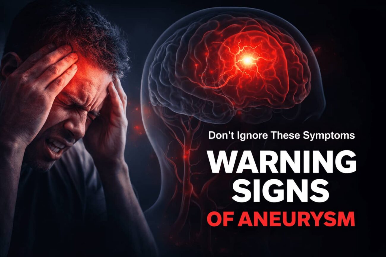
It started with the worst headache of her life. Not a dull throb — an explosion. Peak intensity in under ten seconds. She had never felt anything like it, and she was right not to ignore it.
Symptoms of an aneurysm are one of medicine’s most consequential warning signs — because an aneurysm that ruptures gives you a very short window to act. Yet for the majority of people living with one, there are no symptoms at all. The bulge forms silently in an artery wall, pressing on nothing, causing no pain, waiting.
That silence is what makes understanding the warning signs so critical. When an aneurysm does signal its presence — through pain, vision changes, or neurological shifts — those signs are specific enough to recognise. When it ruptures, the signs are unmistakable. And in both cases, your response in the first minutes determines the outcome.
This guide covers every type of aneurysm, every warning sign, the differences in how symptoms present in women, and exactly when to call emergency services — based on current medical understanding.
What Is an Aneurysm and Why Is It So Dangerous?
An aneurysm is a localised bulge or ballooning in the wall of a blood vessel, caused by a weakness in the arterial wall. As blood continuously flows through the vessel under pressure, the weakened section stretches outward — like a worn spot on a tyre that forms a bubble under pressure.
The danger lies in two distinct phases. In the unruptured phase, a large or growing aneurysm can compress surrounding brain tissue, nerves, or adjacent structures — producing neurological symptoms. In the rupture phase, the wall tears open, releasing blood in ways that can destroy brain tissue within minutes, cause catastrophic drops in blood pressure, or cut off oxygen supply to critical organs.
Aneurysms can form in virtually any artery in the body. The two most medically significant locations are the brain (cerebral or intracranial aneurysm) and the aorta — the body’s largest artery, running from the heart through the chest and abdomen.
Cerebral (Brain / Intracranial) Aneurysm
Forms in the arteries supplying blood to the brain, most commonly at branching points along the Circle of Willis. Often called a “berry aneurysm” due to its rounded shape. Rupture causes subarachnoid hemorrhage — bleeding into the space surrounding the brain. The most immediately life-threatening type.
Thoracic Aortic Aneurysm (TAA)
Forms in the section of the aorta running through the chest. Often associated with connective tissue disorders (Marfan syndrome, Ehlers-Danlos) or bicuspid aortic valve. Can compress the trachea or oesophagus before rupture, causing breathing or swallowing difficulties.
Abdominal Aortic Aneurysm (AAA)
The most common aortic aneurysm — forms in the section of the aorta below the diaphragm. Strongly associated with smoking and atherosclerosis. Often completely silent until rupture. Routine screening is recommended for men over 65 who have ever smoked.
Peripheral Aneurysm
Can form in the popliteal artery (behind the knee), femoral artery (groin), or carotid artery (neck). Less immediately life-threatening than brain or aortic aneurysms, but can cause limb ischemia, stroke, or embolism if untreated.
Symptoms of an Aneurysm: The Warning Signs You Need to Know
Symptoms of an aneurysm depend on its location and whether it has ruptured. An unruptured brain aneurysm may cause pain behind one eye, a drooping eyelid, double vision, or a dilated pupil. A ruptured brain aneurysm causes a sudden, explosive headache — the worst of one’s life — often followed by neck stiffness, vomiting, and loss of consciousness. Most unruptured aneurysms produce no symptoms at all.
Understanding the signs of an aneurysm brain before rupture requires knowing that symptoms arise from physical compression, not from bleeding. A large aneurysm growing near the oculomotor nerve (cranial nerve III), for example, will compress it — producing the characteristic drooping eyelid and fixed, dilated pupil long before the vessel wall gives way.
Unruptured Brain Aneurysm — Warning Signs
Pain Behind or Above One Eye
A persistent, localised ache or pressure above or behind one eye — often on the same side as the aneurysm. Distinct from typical headache in its focal nature and resistance to over-the-counter pain relief.
Drooping Eyelid (Ptosis)
A hallmark sign of compression on the oculomotor nerve. The affected eyelid droops or partially closes. This is a serious neurological warning sign that should never be attributed to fatigue alone.
Double or Blurred Vision
Compression of cranial nerves III, IV, or VI disrupts coordinated eye movement, producing double vision (diplopia). New or unexplained visual disturbances always warrant investigation.
Dilated, Unresponsive Pupil
One pupil that is notably larger than the other and does not constrict in response to light — a sign of oculomotor nerve compression. This is a medical emergency on its own.
Facial Numbness or Tingling
One-sided numbness, tingling, or weakness of the face can indicate an aneurysm pressing on trigeminal nerve branches or adjacent cortical areas.
Localised, Persistent Headaches
New, recurring headaches in a specific region of the head — particularly those that have changed in character, intensity, or frequency — should always prompt evaluation in at-risk individuals.
Signs of a Brain Aneurysm (Intracranial Aneurysm) in Detail
The brain relies on a constant, precisely regulated blood supply delivered through the arterial network known as the Circle of Willis. Aneurysms preferentially form at the bifurcation points of these arteries — where haemodynamic stress is greatest and the vessel wall experiences the highest turbulence.
The symptoms of intracranial aneurysm before rupture are neurological in character because the expanding vessel compresses adjacent brain tissue, cranial nerves, or the optic chiasm. The specific symptoms that appear depend entirely on where in the brain the aneurysm is located.
Common signs of an unruptured brain aneurysm include:
- Persistent pain above or behind one eye, often on the side of the aneurysm
- A drooping eyelid (ptosis) on one side
- Double vision or partial loss of vision
- One pupil that is dilated and unresponsive to light
- Numbness, tingling, or weakness on one side of the face
- New or worsening localised headaches, distinct from previous patterns
- Stiff neck without infection or injury
- Short-term memory lapses or difficulty concentrating (rare, large aneurysms)
It is critical to note: the absence of these symptoms does not mean an aneurysm is not present. The majority of brain aneurysms are discovered incidentally — during brain scans performed for entirely unrelated reasons. If you have known risk factors (family history, connective tissue disorder, polycystic kidney disease), discuss proactive screening with your doctor even in the absence of symptoms.
Symptoms of Brain Aneurysm in Women: What’s Different
Women are diagnosed with brain aneurysms at roughly twice the rate of men — and face a higher lifetime risk of subarachnoid haemorrhage from rupture. The reasons are not fully understood, but hormonal factors are strongly implicated. The decline in oestrogen during and after menopause is thought to compromise arterial wall integrity, making women over 50 particularly vulnerable.
The core symptoms of brain aneurysm in women are the same as in men, but several clinically important differences exist in how those symptoms present and how they are interpreted:
🚺 Brain Aneurysm in Women
- Migraine-type headaches may mask or delay recognition of aneurysm pain
- Hormonal changes (menopause, pregnancy) increase rupture risk
- More likely to attribute vision changes or neck stiffness to hormonal causes
- Higher lifetime incidence of subarachnoid haemorrhage than men
- Pregnancy-related vascular stress raises risk in the third trimester and postpartum
- Postmenopausal women with new, focal headaches warrant proactive evaluation
🚹 Brain Aneurysm in Men
- Slightly lower overall incidence but similar rupture mortality
- More commonly linked to smoking and hypertension as primary drivers
- Sudden-onset severe headache more likely to prompt immediate emergency response
- Lower baseline migraine frequency reduces diagnostic ambiguity
- Uric acid and atherosclerosis-related vascular changes contribute more often
- Higher rates of abdominal aortic aneurysm (AAA) than women
The pattern of women receiving delayed diagnosis for vascular conditions is well-documented. Just as kidney stone symptoms in women are frequently misattributed to gynaecological causes, neurological symptoms in women are sometimes too readily attributed to migraine or anxiety — delaying the investigation that could confirm or rule out an intracranial aneurysm.
If you are a woman over 40 with a first-degree relative who had a brain aneurysm, and you develop new or changing headache patterns, do not self-manage. Request evaluation.
Signs of an Aortic Aneurysm: Chest and Abdominal Warning Signs
Aortic aneurysms behave very differently from brain aneurysms. They grow slowly — often over decades — and rarely cause noticeable symptoms until they are very large or have ruptured. When symptoms do appear, they reflect the physical compression of surrounding structures by the enlarging vessel.
Signs of an Aortic Aneurysm (Abdominal — AAA)
- A pulsating, throbbing sensation in the abdomen — like feeling a heartbeat in your belly
- Deep, persistent aching in the abdomen, lower back, or flanks
- Pain that radiates into the groin or upper thighs
- A persistent sense of abdominal fullness or pressure
- Back pain that is constant, not postural, and does not respond to rest
Signs of a Thoracic Aortic Aneurysm (TAA)
- Deep, aching pain in the chest, jaw, neck, or upper back
- Shortness of breath or difficulty breathing
- Difficulty swallowing (compression of the oesophagus)
- Hoarseness without respiratory illness (compression of the recurrent laryngeal nerve)
- A harsh, high-pitched breathing sound (stridor)
Most small aortic aneurysms — those under 4cm — are found incidentally during imaging for other conditions. Aneurysms above 5.5cm carry significantly elevated rupture risk and typically require surgical or endovascular repair. Between 4–5.5cm, regular surveillance imaging (every 6–12 months) is standard.
Interestingly, nutritional health intersects with vascular integrity. Magnesium, for instance, plays a direct role in regulating arterial smooth muscle tone and systemic blood pressure — a key risk factor for aneurysm development. If you suspect nutritional gaps may be affecting your vascular health, our guide on symptoms of magnesium deficiency covers 12 clinical warning signs worth reviewing.
Signs of an Aneurysm Rupture: A Medical Emergency
A ruptured brain aneurysm is among the most time-critical medical emergencies in existence. Blood released into the subarachnoid space — the area between the brain and the surrounding membranes — causes an immediate, catastrophic rise in intracranial pressure. Irreversible brain damage begins within minutes.
- A sudden, explosive headache — the most severe headache of your life, peaking within seconds (the “thunderclap headache”)
- Loss of consciousness, even briefly
- Sudden confusion, disorientation, or inability to speak
- Stiff neck combined with headache and light sensitivity
- Seizure with no prior history
- Sudden vision loss, drooping face, or one-sided weakness
- Sudden, tearing chest or back pain (possible aortic rupture)
Do not drive. Do not wait to see if it improves. Call emergency services immediately.
The Thunderclap Headache — The Most Important Warning Sign
The thunderclap headache is the defining symptom of a ruptured brain aneurysm. People consistently describe it the same way: an explosion in the head, maximum intensity reached in seconds, unlike anything experienced before. It is not gradually worsening — it is full-intensity almost instantly.
Approximately 10–15% of people with a subarachnoid haemorrhage die before reaching the hospital. Of those who reach emergency care, outcomes depend almost entirely on the speed and quality of treatment. Every minute of delay increases the likelihood of rebleeding, vasospasm (secondary arterial constriction), and permanent neurological damage.
Signs of a Ruptured Aortic Aneurysm
A ruptured abdominal or thoracic aortic aneurysm presents differently from a brain aneurysm rupture, but is equally — often more rapidly — fatal without emergency surgery:
- Sudden, severe, tearing pain in the abdomen, chest, or back
- Rapid drop in blood pressure (haemorrhagic shock): pallor, clamminess, confusion
- Rapid, weak heartbeat
- Pulsating abdominal mass that may be visible or palpable
- Loss of consciousness
Ruptured aortic aneurysms have an overall mortality of up to 80% — most deaths occur before the patient reaches hospital. The only intervention that offers survival is emergency open or endovascular surgery.
Aneurysm vs. Stroke: How to Tell the Difference
The symptoms of a ruptured brain aneurysm overlap with those of a stroke — both can cause sudden headache, facial drooping, slurred speech, and confusion. The distinction matters clinically because treatment pathways differ, but in either case the emergency response is identical: call for help immediately.
| Feature | Ruptured Brain Aneurysm | Ischaemic Stroke |
|---|---|---|
| Cause | Blood vessel bursts, bleeding into brain | Blood vessel blocked, no blood flow to area |
| Headache | Sudden, explosive, worst ever (thunderclap) | Usually absent or mild |
| Onset Speed | Immediate — peak intensity within seconds | Often gradual, or sudden without headache |
| Loss of Consciousness | Common, may occur within minutes | Less common in initial presentation |
| Neck Stiffness | Often present (meningismus) | Usually absent |
| Light Sensitivity | Frequently present | Rare |
| Treatment Approach | Surgical clipping or endovascular coiling | Clot-busting drugs (tPA) or thrombectomy |
Who Is Most at Risk for Developing an Aneurysm?
Several factors — both genetic and lifestyle-related — significantly raise the risk of aneurysm formation and rupture:
- 1High blood pressure (hypertension) — The single most significant modifiable risk factor. Chronic elevated blood pressure exerts constant excess stress on arterial walls, gradually weakening them over time. Controlling blood pressure to below 120/80 mmHg is the most impactful preventive step available.
- 2Smoking — Directly damages arterial wall connective tissue, accelerates atherosclerosis, and raises blood pressure. Smokers have 2–3x the rupture risk of non-smokers. The relationship is dose-dependent: the longer and heavier the smoking history, the higher the risk.
- 3Family history — Having one first-degree relative (parent, sibling) with a brain aneurysm roughly doubles your risk. Having two first-degree relatives raises it further — to a level that typically justifies screening with MRA (magnetic resonance angiography).
- 4Genetic connective tissue disorders — Polycystic kidney disease, Marfan syndrome, Ehlers-Danlos syndrome (vascular type), and neurofibromatosis are all independently associated with aneurysm formation due to inherent arterial wall weakness.
- 5Sex and hormonal status — Women over 50 (post-menopause) have a disproportionately higher rate of brain aneurysm and subarachnoid haemorrhage. The oestrogen decline associated with menopause is the likely mechanism.
- 6Cocaine and stimulant use — Cause acute, dramatic spikes in blood pressure that can trigger rupture of pre-existing aneurysms. Cocaine use is one of the most common precipitating triggers of subarachnoid haemorrhage in adults under 40.
How Aneurysms Are Diagnosed: Imaging and Tests Explained
When a doctor suspects an aneurysm based on symptoms, risk profile, or incidental findings, several imaging modalities can confirm the diagnosis. The choice depends on clinical urgency, available resources, and whether rupture has occurred.
| Imaging Type | What It Shows | Best Used For | Limitations |
|---|---|---|---|
| CT Scan (non-contrast) | Blood in the brain after rupture; intracranial haemorrhage | First-line in emergency settings; fastest way to confirm rupture | May miss very small or unruptured aneurysms |
| CT Angiography (CTA) | Precise aneurysm size, location, shape, and surrounding vessels | Pre-surgical planning; detailed anatomy of unruptured aneurysm | Radiation + contrast dye; not ideal in renal impairment |
| MRI / MRA | Detailed brain structure; aneurysm detection without radiation | Screening in high-risk individuals; follow-up of known aneurysms | Takes longer; may miss acute bleeding vs. CT |
| Cerebral Angiography (DSA) | Gold standard — precise vessel anatomy, aneurysm neck, flow patterns | Pre-treatment planning; when other imaging is inconclusive | Invasive; small procedural stroke risk (<0.5%) |
| Lumbar Puncture (LP) | Blood or xanthochromia in cerebrospinal fluid after rupture | When CT is negative but thunderclap headache persists (>6 hours) | Invasive; results can be ambiguous in first 2 hours |
| Abdominal Ultrasound | AAA size and shape; serial monitoring | Screening for abdominal aortic aneurysm; low cost and no radiation | Poor for thoracic or intracranial aneurysms |
Treatment Options: From Watchful Waiting to Surgery
Not every aneurysm requires immediate surgery. Treatment decisions are individualised, weighing the rupture risk of the aneurysm against the procedural risks of intervention.
For Unruptured Aneurysms
- Watchful waiting and surveillance: Small aneurysms (under 5mm) in low-risk locations with no symptoms are often monitored with periodic MRA every 1–3 years. Lifestyle modification (blood pressure control, smoking cessation) is mandatory alongside monitoring.
- Surgical clipping: A neurosurgeon opens the skull and places a tiny metal clip at the base of the aneurysm, cutting off blood flow into it. A durable, long-established procedure with permanent results.
- Endovascular coiling (EVOH): A catheter is guided through an artery to the aneurysm site, where tiny platinum coils are deployed inside the aneurysm, causing it to clot and seal off. Less invasive than open surgery; now the preferred method in many centres.
- Flow-diverting stents: A mesh device placed in the parent artery redirects blood flow away from the aneurysm, gradually causing it to shrink. Used for large or fusiform aneurysms not amenable to coiling.
For Ruptured Aneurysms
Emergency intervention — either surgical clipping or endovascular coiling — is required as rapidly as possible. Intensive care management addresses raised intracranial pressure, vasospasm (secondary arterial narrowing that causes delayed ischaemic injury), hydrocephalus, and systemic complications.
For evidence-based clinical guidelines on aneurysm management, the American Heart Association provides comprehensive cardiovascular guidance, and the Mayo Clinic’s brain aneurysm overview offers clear, patient-facing information on diagnosis and treatment pathways.
Frequently Asked Questions
Most aneurysms produce no symptoms at all before they rupture or grow large enough to compress nearby structures. When early symptoms do occur, they typically include a pain above or behind one eye, a drooping eyelid, double vision, or a dilated pupil. A new, persistent, or worsening headache in a specific location — especially in someone with known risk factors — also warrants investigation.
A brain aneurysm headache — associated with rupture — is described as the worst headache of one’s life, reaching peak intensity within seconds rather than minutes. It is often called a “thunderclap headache.” Before rupture, some aneurysms cause a dull, localised ache above or behind one eye, distinct from typical headache in its persistence and focal nature. Any sudden, explosive, unprecedented headache should be treated as a medical emergency until proven otherwise.
Yes. Women are diagnosed with brain aneurysms at approximately twice the rate of men, and have a higher lifetime risk of subarachnoid haemorrhage. The hormonal changes associated with menopause — specifically the decline in oestrogen — are thought to weaken arterial walls, increasing aneurysm risk. Women over 50 with risk factors such as hypertension, smoking, or family history should discuss proactive brain aneurysm screening with their doctor.
The primary sign of a ruptured brain aneurysm is a sudden, explosive “thunderclap” headache — the worst of one’s life. Additional signs include neck stiffness, nausea and vomiting, light sensitivity, confusion, seizure, loss of consciousness, and one-sided facial or body weakness. A ruptured aortic aneurysm causes sudden, tearing chest or abdominal pain with haemorrhagic shock. Both are life-threatening emergencies requiring immediate emergency services.
No. Aneurysms do not resolve or shrink on their own. Small, unruptured aneurysms may remain stable for years and are monitored rather than immediately treated. However, the aneurysm itself does not disappear — it requires ongoing surveillance and lifestyle management. The decision between continued monitoring and surgical intervention is made based on aneurysm size, location, growth rate, and individual patient risk factors.
A brain aneurysm rupture causes a haemorrhagic event — blood escapes into the brain or surrounding space, causing pressure damage. An ischaemic stroke occurs when a blood clot blocks an artery, cutting off blood supply. Both share some symptoms — sudden weakness, speech difficulty, confusion — but a thunderclap headache is strongly associated with aneurysm rupture and is usually absent in ischaemic stroke. Both require emergency treatment. Do not attempt to determine which condition is occurring — call for emergency help immediately.
Proactive screening is recommended for people with two or more first-degree relatives with brain aneurysms, those with polycystic kidney disease, Marfan syndrome, or Ehlers-Danlos syndrome (vascular type), and individuals with a prior history of aneurysm. Screening is typically performed with MRA (magnetic resonance angiography) — no radiation, no invasive procedures. If you have one of these risk factors and have never been screened, discuss it with your GP or neurologist.
Summary: Recognising Aneurysm Symptoms Can Save Your Life
Symptoms of an aneurysm — whether in the brain, chest, or abdomen — are among the most important warning signs in medicine. They are also among the most frequently missed, because most aneurysms are silent until a critical moment arrives.
Key points to remember:
- Most aneurysms produce no symptoms before they grow large or rupture
- Signs of a brain aneurysm include pain behind one eye, a drooping eyelid, double vision, and a fixed, dilated pupil — caused by nerve compression, not bleeding
- The symptoms of intracranial aneurysm rupture are unmistakable: a thunderclap headache, neck stiffness, light sensitivity, and often loss of consciousness
- Women face a higher lifetime risk of brain aneurysm and subarachnoid haemorrhage — particularly after menopause — and should not dismiss new neurological symptoms
- Signs of an aortic aneurysm include abdominal pulsation, persistent back pain, and sudden tearing chest or abdominal pain if ruptured
- High blood pressure and smoking are the two most impactful modifiable risk factors — controlling both significantly reduces aneurysm risk
- If you experience a sudden, explosive, unprecedented headache — call emergency services immediately. Do not wait.
Prevention starts with knowing your risk. If you have a family history of aneurysm, a connective tissue disorder, or poorly controlled blood pressure, speak to your doctor about proactive screening. An MRA scan is non-invasive, radiation-free, and could identify an aneurysm long before it causes a crisis.
Disclaimer: This article is for informational and educational purposes only and does not constitute medical advice. Always consult a qualified healthcare professional for diagnosis and treatment of any medical condition, including aneurysm symptoms. In any suspected medical emergency, call emergency services immediately. Read full disclaimer →